Dynamic Renography (⁹⁹ᵐTc-MAG3)
Dynamic renography with ⁹⁹ᵐTc-MAG3 images renal perfusion, uptake and drainage over time. Its key uses are distinguishing obstruction from non-obstructive dilatation (with a furosemide diuretic challenge), quantifying split (differential) function, and assessing renal transplants (perfusion, drainage, ATN vs obstruction vs leak).
MAG3 is highly extracted by the renal tubules, giving good images even when function is impaired. The renogram curve (rise, peak, washout) and the post-diuretic washout half-time (t½) form the basis of interpretation.
When to image
- Hydronephrosis — is it obstructed? (diuretic renography).
- Quantifying differential function before surgery.
- Transplant dysfunction — perfusion, drainage, ATN vs obstruction vs urine leak.
- Assessing drainage after pyeloplasty/stenting.
How to read it
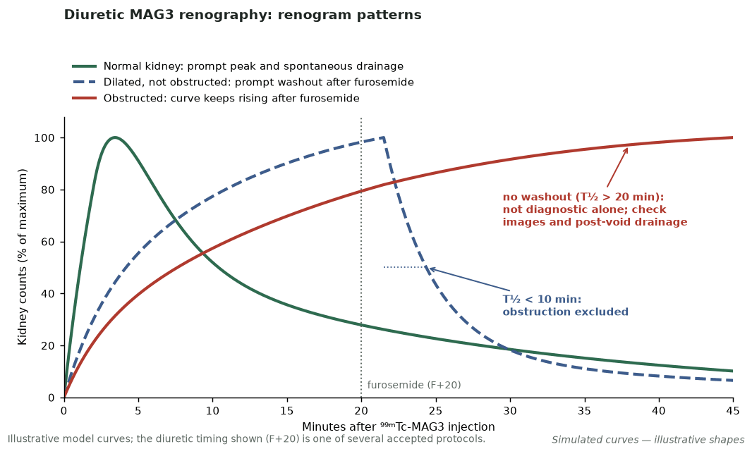
- A post-furosemide T½ <10 min reliably excludes obstruction; a prolonged T½ (classically >20 min) is not diagnostic on its own — interpret with the images, curves and gravity-assisted post-void drainage (e.g. post-void/maximum count ratio).
- Relative uptake gives split function.
- Transplant ATN: relatively preserved perfusion with poor function and parenchymal retention (reduced perfusion favours rejection or vascular compromise); a leak shows tracer accumulating outside the urinary tract (a urinoma may be photopenic early) — SPECT/CT localises it.
Protocol
- Hydrate (oral 5–10 mL/kg 30–60 min before; avoid fasting); catheterise if voiding is difficult or with a neobladder/urinary diversion.
- Dynamic acquisition with furosemide 40 mg (0.5 mg/kg; ~80 mg if function is reduced); timing (F−15, F0, F+20 etc.) has no consensus, but a post-void image is essential.
- Generate renogram curves and split-function values.
Pitfalls
- Dehydration and poor function blunt the diuretic response (false “obstruction”).
- A full bladder/reflux distorts drainage; a markedly dilated pelvis can drain slowly without obstruction (“reservoir” effect).
- Interpret the curve with the clinical question.
Evidence & guidelines
- SNMMI/EANM diuretic renal scintigraphy guideline (Taylor et al., Semin Nucl Med 2018;48:377–390) and adult renal scintigraphy guideline (Blaufox et al., Eur J Nucl Med Mol Imaging 2018;45:2218–2228).
- MAG3 is preferred over DTPA in impaired function.
In depth
- ⁹⁹ᵐTc-MAG3 is highly protein-bound (about 90%), so it is cleared almost entirely by proximal tubular secretion rather than filtration, with an extraction fraction of 40–50%; over 95% leaves the body by 3 h in normal renal function, and 37–185 MBq (1–5 mCi) is adequate for most adult studies.
- ⁹⁹ᵐTc-DTPA is cleared purely by glomerular filtration (about 5–10% protein-bound, so clearance is slightly below inulin) and can measure GFR; its extraction fraction is about 20%, versus 40–50% for MAG3.
- The renogram has three phases (vascular arrival over the first ~30–60 s, parenchymal uptake, and excretion); normal MAG3 and DTPA renograms peak by about 5 min and fall to half-peak by about 15 min.
- Prolonged parenchymal transit (renal-artery stenosis, dehydration, ATN, nephrotoxic drugs) delays and blunts the peak; obstruction produces a continuously rising or flat excretory third segment, and pelvic retention can mimic this.
- After furosemide, a dilated but unobstructed collecting system drains promptly, whereas a true (mechanical) obstruction shows little or no washout; interpretation is unreliable when the kidney's function is poor.
- Because it is secreted by the tubules with higher extraction, MAG3 gives better kidney images than DTPA when GFR is severely reduced, such as in ATN, obstruction or renal failure.
- ⁹⁹ᵐTc-EC (ethylenedicysteine) is a tubular agent with slightly higher clearance than MAG3 and lower hepatic uptake, giving comparable renogram quality and split function.
- Camera-based MAG3 clearance uses background-subtracted, depth- or attenuation-corrected renal counts in an early window (for example 1–2.5 min) as a percentage of injected dose, converted by regression to clearance and normalised to body surface area, without blood or urine sampling; it is less accurate than plasma-sampling methods.
Sources: Taylor, J Nucl Med 2014 · 55:608-15 (PMID 24549283) · SNMMI/EANM renal scintigraphy guideline 2018 (PMID 30167801) · Taylor, J Nucl Med 2014 (PMID 24549283) · 55:786-98 (PMID 24591488) · SNMMI/EANM diuretic renal scintigraphy guideline 2018 (PMID 29852947) · Jain et al. Nucl Med Commun 2018 (PMID 30169343) · Inoue et al. J Nucl Med 1999
- Antenatal hydronephrosis in a 3-month-old boy · Intermediate