Hepatobiliary and Liver–Spleen Imaging
At a glance
- HIDA. ⁹⁹ᵐTc-mebrofenin shows hepatocyte function, bile flow and gallbladder filling in one study.
- Acute cholecystitis. Gallbladder not seen at 3–4 h, or 30 min after morphine, is diagnostic; sensitivity 96%, specificity 90%.
- GBEF. Sincalide 0.02 µg/kg infused over 60 min; normal gallbladder ejection fraction ≥38%.
- Neonatal cholestasis. No bowel activity by 24 h after phenobarbital suggests biliary atresia; bowel activity excludes it.
- Leaks and obstruction. HIDA proves whether a collection is bile and can show obstruction before ducts dilate.
- Other liver agents. Sulfur colloid (Kupffer cells), labelled RBCs (haemangioma) and hepatic-artery MAA (before radioembolisation).
1. Anatomy in brief
- The liver bud arises from the foregut; its cranial part forms the liver, its caudal part the gallbladder and cystic duct.
- Physiological lobes: divided by Cantlie's line (gallbladder fossa to IVC), along the middle hepatic vein. The caudate and quadrate lobes are anatomically right but functionally left.
- Couinaud segments: hepatic veins divide the liver vertically, portal branches horizontally, giving eight segments (I–VIII).
- Caudate lobe (I): drains directly into the IVC, so it is often spared in Budd–Chiari syndrome and appears relatively 'hot' on colloid imaging.
- Gallbladder: 7–10 cm long, 40–50 mL; stores and concentrates bile between meals.
2. Liver–spleen colloid imaging
- ⁹⁹ᵐTc-sulfur colloid: particles of ~0.1–1 µm are phagocytosed by reticuloendothelial cells. Most go to the liver (Kupffer cells), the rest to spleen and bone marrow.
- Colloid shift: in diffuse liver disease or portal hypertension, relatively more tracer goes to spleen and marrow.
- Most focal lesions, benign or malignant, are photopenic because they lack Kupffer cells.
- Historical: ¹⁹⁸Au colloid (411 keV γ, T½ 2.7 days, β⁻ dose) is obsolete.
| Focal 'hot' spot on colloid imaging | Why |
|---|---|
| Focal nodular hyperplasia | Contains Kupffer cells, so uptake is often normal and occasionally increased |
| Budd–Chiari syndrome | Caudate lobe drains separately into the IVC and is spared |
| Superior vena cava obstruction (arm injection) | Collaterals deliver tracer to segment IV (quadrate region) |
| Inferior vena cava obstruction (leg injection) | Collateral flow to the liver |
| Regenerating nodule in cirrhosis | Functioning Kupffer cells |
3. Other liver agents
- ⁹⁹ᵐTc-labelled RBCs (haemangioma): normal arterial flow, reduced activity on early blood-pool images, then increased activity on 1–2 h delayed images, equal to cardiac blood pool. Large haemangiomas may have cold areas from thrombosis or fibrosis. SPECT improves detection.
- ⁹⁹ᵐTc-MAA hepatic-artery perfusion: particles of 10–90 µm lodge in arterioles on first pass. Used before radioembolisation to show tumour and extrahepatic perfusion and to measure lung shunting. A lung shunt above 20% contraindicates resin microspheres; the lung dose is kept below 30 Gy (50 Gy cumulative).
- ¹¹¹In-pentetreotide: somatostatin-receptor imaging of neuroendocrine liver metastases (111 MBq planar, 222 MBq SPECT); now largely replaced by ⁶⁸Ga-DOTA-peptide PET.
- ⁶⁷Ga citrate and labelled leukocytes: historical use for tumour and abscess; FDG PET/CT has largely replaced them.
4. Hepatobiliary scintigraphy (HIDA)
The tracers
- ⁹⁹ᵐTc-iminodiacetic acid (IDA) derivatives, originally lidocaine analogues; the ring substitutions change their kinetics.
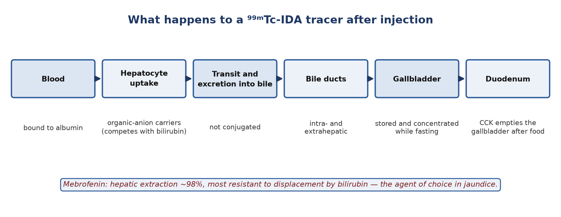
- Mebrofenin (bromo-trimethyl IDA): hepatic extraction ~98%; the most resistant to displacement by high bilirubin.
- Disofenin (di-isopropyl IDA): hepatic extraction ~89%.
- They are transported bound to albumin, taken up by hepatocytes through organic-anion pathways (competing with bilirubin) and excreted into bile without conjugation.
Preparation and protocol
| Step | Recommendation (SNM guideline 4.0) |
|---|---|
| Fasting | At least 2 h, preferably 6 h (a meal in the previous hours makes the gallbladder contract) |
| Fasted >24 h or on parenteral nutrition | The gallbladder may be full of viscous bile: pretreat with sincalide 0.02 µg/kg over 30–60 min, 15–30 min before the tracer |
| Opioids | Contract the sphincter of Oddi and mimic obstruction: delay the study for about 4 half-lives of the drug |
| Activity (adult) | 111–185 MBq; more if bilirubin is high |
| Activity (child) | 1.8 MBq/kg, minimum 18.5 MBq (37 MBq in neonates with hyperbilirubinaemia) |
| Imaging | Dynamic/sequential anterior images for 60 min; right lateral or LAO views separate gallbladder from duodenum; delayed images to 3–4 h (sometimes 18–24 h) |
Reading the study
- Flow: spleen and kidneys appear first; the liver appears later because most of its supply is portal. Increased flow to the gallbladder fossa can accompany severe acute cholecystitis.
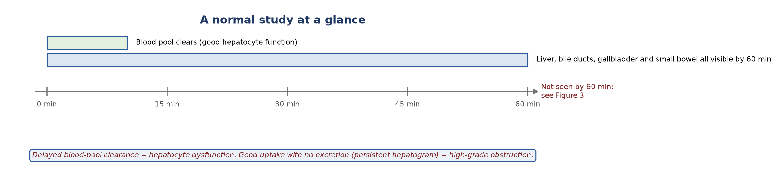
- Hepatocyte function: judged by how fast the cardiac blood pool clears (normally within minutes).
- Gallbladder: normally fills within 60 min; filling beyond 60 min is delayed.
- Ducts and bowel: ducts and small bowel should be seen within 60 min. Scintigraphy shows whether ducts drain, not whether they are dilated.
5. Clinical uses
Acute cholecystitis
- Usually caused by a stone obstructing the cystic duct.
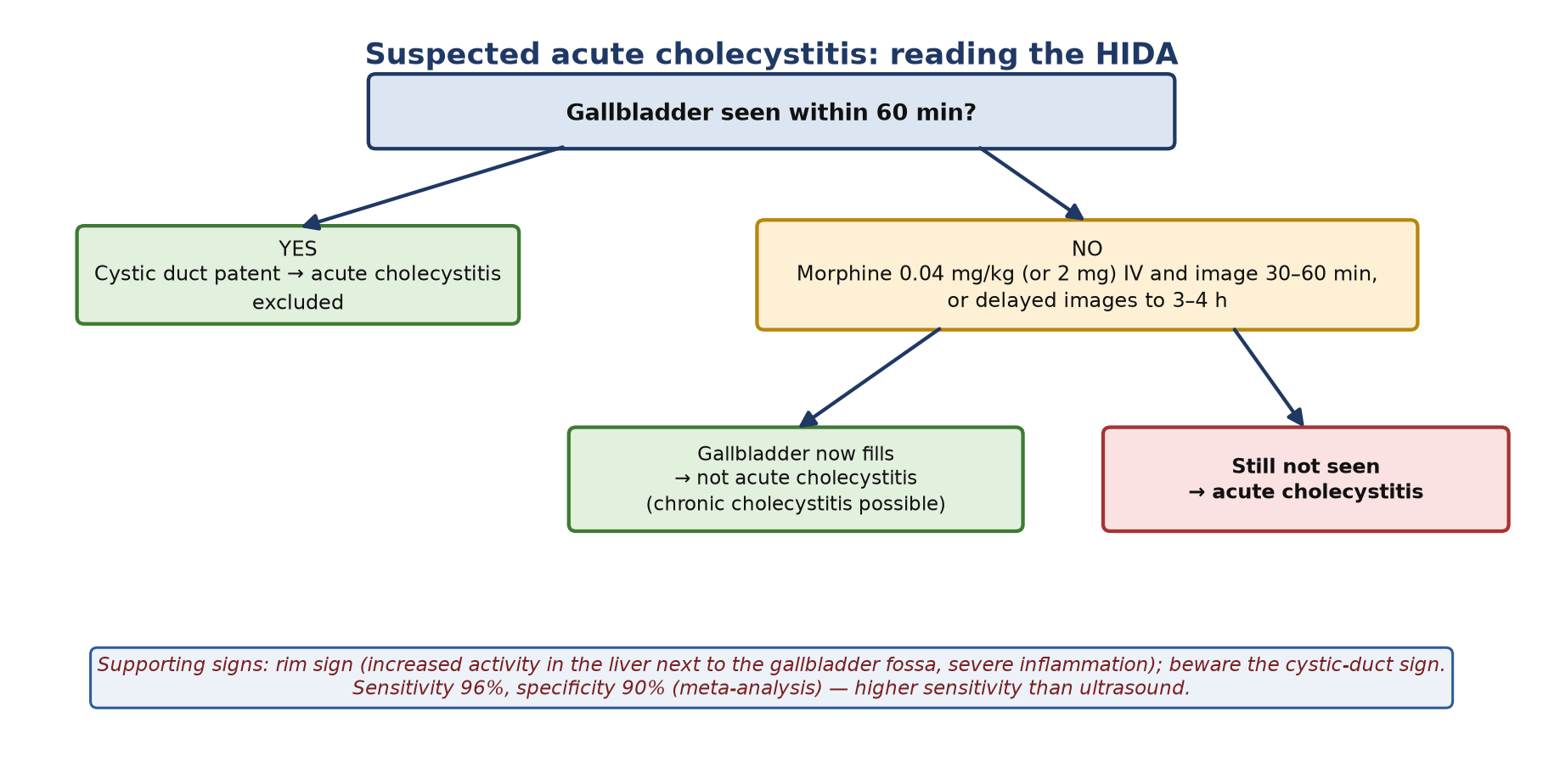
- Non-filling at 60 min is abnormal but not yet diagnostic. Persistent non-visualisation at 3–4 h, or 30 min after morphine, is diagnostic (calculous and acalculous).
- Morphine augmentation: 0.04 mg/kg (or 2 mg) IV over 2–3 min contracts the sphincter of Oddi, raises duct pressure and fills the gallbladder if the cystic duct is patent; image a further 30–60 min.
- Accuracy: sensitivity 96% and specificity 90% in a meta-analysis of 57 studies, higher sensitivity than ultrasound (81%).
- Rim sign: increased pericholecystic hepatic activity, a marker of severe inflammation (gangrene, perforation risk). Cystic-duct sign: a small focus of dilated cystic duct mistaken for the gallbladder.
Chronic cholecystitis and gallbladder ejection fraction
- Delayed filling (>60 min) is common, but filling may be normal.
- Standard GBEF protocol: after gallbladder filling, sincalide 0.02 µg/kg infused over 60 min; GBEF at 60 min. Normal ≥38% (interdisciplinary consensus).
- A low GBEF supports chronic cholecystitis or functional gallbladder disorder in the right clinical setting; a normal GBEF argues against it.
Biliary atresia
- Progressive fibro-obliterative cholangiopathy of infancy; Kasai portoenterostomy is most effective when done early (ideally within the first 60 days).
- A gallbladder on ultrasound does not exclude atresia.
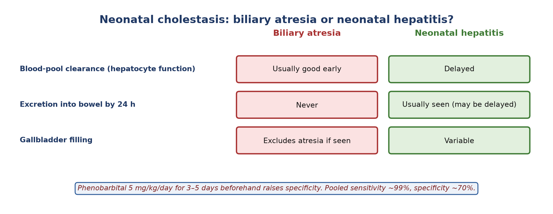
- Protocol: phenobarbital 5 mg/kg/day for 3–5 days to stimulate bile flow; image to 24 h.
- Pattern: atresia — good early uptake but no bowel activity by 24 h; neonatal hepatitis — poor uptake but bowel activity usually appears. Pooled sensitivity 98.7%, specificity 70.4%.
Bile leak
- After cholecystectomy, trauma or biliary surgery. Ultrasound and CT show fluid; HIDA shows whether it is bile.
- A progressively enlarging collection in the gallbladder fossa or hepatic hilum, spreading over the liver dome, into the gutters or free in the abdomen. Image drains and collection bags; right lateral decubitus views help.
Biliary obstruction
- High-grade (recent) obstruction: good hepatocyte uptake with no excretion (persistent hepatogram), often before ducts dilate on ultrasound (which can take 24–72 h).
- Partial obstruction: prompt uptake and excretion but delayed ductal clearance, retained or increasing ductal activity and delayed biliary-to-bowel transit.
- Sphincter of Oddi dysfunction: after cholecystectomy, a partial obstruction at the sphincter without stone, stricture or tumour; scintigraphic scoring systems help select patients.
- Choledochal cyst: a non-obstructed cyst fills slowly and retains tracer; this confirms its connection to the biliary tree.
- Biliary-enteric anastomoses (e.g. Roux loop, Whipple): HIDA shows leak, patency and recurrent obstruction; bowel activity by 60 min supports patency.
- Other uses: biliary stent function, enterogastric bile reflux, focal nodular hyperplasia (normal or increased uptake with delayed clearance).
6. Spleen and red cells
- The spleen is seen with colloids, labelled red cells (including heat-damaged RBCs for splenic tissue), leukocytes and somatostatin-receptor tracers, but not with HIDA agents.
- ⁵¹Cr-labelled red cells: hexavalent ⁵¹Cr enters the cell, is reduced to trivalent chromium and binds haemoglobin; released chromium does not relabel other cells. Used for red-cell volume and survival (with organ counting), not imaging.
Summary
- Fast 2–6 h; pretreat with sincalide after >24 h fasting; delay the study for ~4 half-lives of an opioid.
- Gallbladder not seen at 3–4 h or 30 min after morphine = acute cholecystitis.
- GBEF: 60-min sincalide infusion, normal ≥38%.
- No bowel activity by 24 h after phenobarbital suggests biliary atresia.
- HIDA identifies bile leaks and shows obstruction before duct dilatation.
- Colloid, RBC and MAA studies answer different liver questions.
Test yourself
5 quick questions. Pick an answer to see the explanation.
1. The gallbladder is not seen at 60 min. After morphine 0.04 mg/kg it fills at 20 min. The diagnosis is:
2. The standard sincalide protocol for gallbladder ejection fraction is:
3. A 6-week-old infant, pretreated with phenobarbital, shows prompt hepatic uptake but no bowel activity at 24 h. Most likely:
4. Which HIDA agent is preferred when bilirubin is markedly raised?
5. On sulfur colloid imaging, the caudate lobe is hot and the rest of the liver patchy. Consider:
References
- Tulchinsky M, Ciak BW, Delbeke D, et al. SNM practice guideline for hepatobiliary scintigraphy 4.0. J Nucl Med Technol. 2010;38(4):210-8.
- Ziessman HA, Tulchinsky M, Lavely WC, et al. Sincalide-stimulated cholescintigraphy: a multicenter investigation to determine optimal infusion methodology and gallbladder ejection fraction normal values. J Nucl Med. 2010;51(2):277-81.
- DiBaise JK, Richmond BK, Ziessman HH, et al. Cholecystokinin-cholescintigraphy in adults: consensus recommendations of an interdisciplinary panel. Clin Gastroenterol Hepatol. 2011;9(5):376-84.
- Kiewiet JJ, Leeuwenburgh MM, Bipat S, et al. A systematic review and meta-analysis of diagnostic performance of imaging in acute cholecystitis. Radiology. 2012;264(3):708-20.
- Kianifar HR, Tehranian S, Shojaei P, et al. Accuracy of hepatobiliary scintigraphy for differentiation of neonatal hepatitis from biliary atresia: systematic review and meta-analysis of the literature. Pediatr Radiol. 2013;43(8):905-19.
Practise with a case