Hepatobiliary Scintigraphy (HIDA)
Snapshot
HIDA (⁹⁹ᵐTc-mebrofenin) images hepatocyte extraction and biliary excretion. Its flagship use is acute cholecystitis: persistent non-visualisation of the gallbladder at 3–4 hours, or 30 minutes after morphine augmentation, is diagnostic (sensitivity ≈96%, specificity ≈90%). It also detects bile leaks and, in neonates, helps distinguish biliary atresia from neonatal hepatitis.
Read the full article →In-depth, fully referenced version
The tracer is extracted by hepatocytes and excreted into bile, so normal images show liver → bile ducts → gallbladder → bowel. A cystic-duct obstruction blocks gallbladder filling — the basis of the cholecystitis diagnosis.
GB non-fillAcute cholecystitis
MorphineAugmentation
NeonatalAtresia vs hepatitis
When to image
- Suspected acute cholecystitis with equivocal ultrasound.
- Suspected bile leak after cholecystectomy or trauma.
- Neonatal cholestasis — biliary atresia vs neonatal hepatitis.
- Assessing biliary-enteric drainage/function.
How to read it
- Gallbladder not seen at 60 min is abnormal but not diagnostic; still not seen at 3–4 h, or 30 min after morphine → acute cholecystitis.
- Tracer outside the biliary tree → bile leak.
- Absent bowel excretion in a neonate (after priming) → possible biliary atresia.
Protocol
- Fast at least 2 h, preferably 6 h; if fasting >24 h, pretreat with sincalide; delay ~4 half-lives after opioids; morphine augmentation (0.04 mg/kg) if non-filling at 60 min.
- Dynamic imaging to 60 min with delayed views as needed.
- Phenobarbitone priming for neonatal atresia studies.
Pitfalls
- Prolonged fasting or TPN causes gallbladder non-filling (false positive) — use morphine/CCK.
- High bilirubin reduces hepatic extraction and image quality.
- Chronic cholecystitis may show delayed filling.
Evidence & guidelines
- Meta-analysis (Kiewiet et al., Radiology 2012;264:708–720): cholescintigraphy sensitivity 96%, specificity 90% for acute cholecystitis — higher sensitivity than ultrasound (81%).
- SNM practice guideline for hepatobiliary scintigraphy 4.0 (Tulchinsky et al., J Nucl Med Technol 2010;38:210–218) covers augmentation and neonatal protocols.
In depth
- Mebrofenin has about 98% hepatic extraction, a hepatic excretion half-time of about 17 min and about 1% renal excretion, compared with 88%, 19 min and about 9% for disofenin, so it is preferred in hyperbilirubinaemia and hepatic dysfunction.
- Adults receive 111–185 MBq (3–5 mCi) of ⁹⁹ᵐTc-mebrofenin or disofenin, with higher activity in hyperbilirubinaemia; children receive 1.8 MBq/kg (0.05 mCi/kg), minimum 18.5 MBq (0.5 mCi), and jaundiced neonates at least 37 MBq (1 mCi) with mebrofenin.
- Adults fast for at least 2 h and preferably 6 h; opioids are withheld for about four half-lives of the drug; if fasting exceeds 24 h or on TPN, give sincalide 0.02 µg/kg over 30–60 min, finishing 15–30 min before tracer injection.
- Dynamic anterior (or LAO) imaging at 1 frame/min for 60 min with a LEAP or high-resolution collimator and a 140 keV photopeak window, with an optional initial flow phase and right-lateral or oblique views as needed to clarify anatomy.
- Normally the liver is seen immediately with rapid blood-pool clearance, followed by the bile ducts, gallbladder and small bowel, all within 60 min; delayed biliary-to-bowel transit beyond 60 min occurs as a normal variant in up to 20%.
- Gallbladder filling excludes acute cholecystitis with high certainty; persistent non-visualisation at 3–4 h, or 30 min after morphine, is the hallmark of acute (calculous or acalculous) cholecystitis.
- Morphine augmentation (gallbladder not seen at 30–60 min, with tracer in the ducts and small bowel): morphine 0.04 mg/kg or 2 mg IV over 2–3 min, then image for a further 30–60 min; contraindicated with morphine allergy, respiratory depression in non-ventilated patients or raised intracranial pressure in children, and relatively in acute pancreatitis.
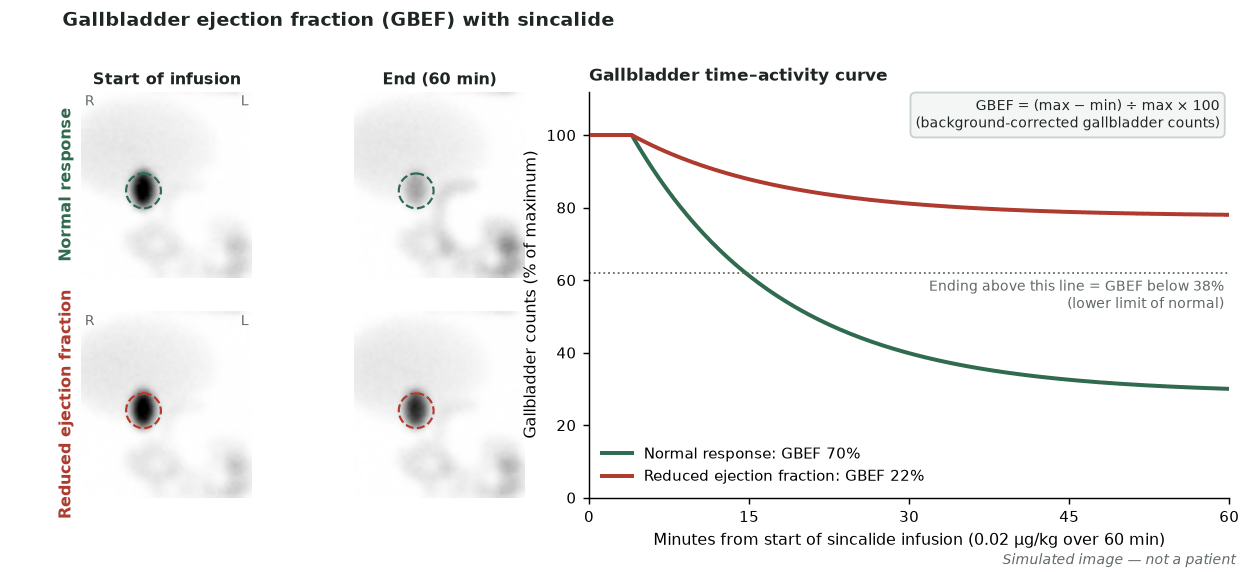
- Gallbladder ejection fraction (35–40° LAO, sincalide 0.02 µg/kg over 60 min): abnormal GBEF <38%, seen in calculous and acalculous disease.
- In biliary atresia there is no tracer in the bowel even on 24-h images, but this is not specific (hepatocellular disease can mimic it); phenobarbitone 5 mg/kg/day for at least 3–5 days, or ursodeoxycholic acid 20 mg/kg/day for 2–3 days, improves specificity, and bowel activity excludes atresia.
- False-positive gallbladder non-filling: insufficient fasting, prolonged fasting/TPN, severe hepatocellular disease, high-grade CBD obstruction, rapid biliary-to-bowel transit, prior cholecystectomy.
Sources: SNMMI hepatobiliary 4.0 · Krishnamurthy data cited in PMC6194760 · SNMMI hepatobiliary 4.0 (Ziessman 2010 normal values)
Practise with a case