Gastric-Emptying Scintigraphy
Snapshot
Gastric-emptying scintigraphy with a standardised ⁹⁹ᵐTc-labelled solid meal is the reference standard for diagnosing gastroparesis (and dumping syndrome). Serial imaging over 4 hours quantifies the percentage of meal retained; delayed emptying supports gastroparesis when correlated with symptoms.
A standardised low-fat egg-white meal is labelled and imaged at set intervals. Standardisation of meal and timing is essential because emptying varies with meal composition.
Standard mealReference test
4-hour imagingSolids
% retainedQuantifies delay
When to image
- Suspected gastroparesis (nausea, early satiety, vomiting) with normal endoscopy.
- Suspected rapid emptying/dumping after gastric surgery.
- Objectively quantifying emptying to guide therapy.
How to read it
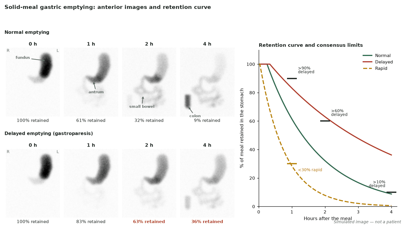
- Report % retention at 1, 2 and 4 h against consensus limits: delayed if >90% at 1 h, >60% at 2 h or >10% at 4 h.
- Delayed emptying beyond reference limits supports gastroparesis.
- Rapid emptying (<30% retention at 1 h) supports dumping.
Protocol
- Standardised ⁹⁹ᵐTc-sulphur-colloid low-fat egg-white meal (with toast, jam and water) eaten within 10 min after an overnight fast (≥6 h).
- Stop prokinetics, opioids and anticholinergics (usually 48–72 h, by half-life); in diabetics record glucose and treat or reschedule if >275 mg/dL (≈15 mmol/L).
- Images at 0, 1, 2 and 4 h (solids); stopping at 2 h misses some delayed emptying.
Pitfalls
- Non-standardised meals/timing invalidate reference ranges.
- Hyperglycaemia slows emptying — control glucose.
- Medications (prokinetics, opioids) alter results.
Evidence & guidelines
- ANMS/SNM consensus (Abell et al., J Nucl Med Technol 2008;36:44–54) defines the standardised solid-meal protocol and normal values.
- Interpret emptying with symptoms and glycaemic status.
In depth
- Standardised low-fat egg-white meal: 120 g liquid egg white (about two large eggs), two slices of white bread, 30 g jam and 120 mL water, labelled with 18.5–37 MBq (0.5–1 mCi) ⁹⁹ᵐTc-sulfur colloid; about 255 kcal.
- Image at 0, 1, 2 and 4 h upright, 1-min anterior + posterior views.
- Quantify gastric counts as the geometric mean (square root of anterior × posterior counts), decay-corrected and normalised to 100% at time 0.
- Normal upper limits of retention: 90% at 1 h, 60% at 2 h, 10% at 4 h; delayed emptying exceeds any of these.
- Rapid emptying = <30% retained at 1 h; a 4-h study detects delayed emptying missed at 2 h.
- Severity by 4-h retention: mild 11–20%, moderate 21–35%, severe 36–50%, very severe >50%.
- In diabetic patients, check glucose before the study and lower it or reschedule if above 275 mg/dL (about 15 mmol/L), because hyperglycaemia delays emptying; stop prokinetics, opioids and anticholinergic antispasmodics at least 2 days (48 h) before.
Sources: SNM/ANMS consensus on gastric emptying scintigraphy, Abell 2008 (PMID 18287197)
Practise with a case