Gastrointestinal Transit Studies
Snapshot
Radionuclide transit studies quantify motility beyond the stomach: oesophageal transit (a labelled swallowed bolus), and small-bowel and colonic transit (a labelled meal followed over hours to days). They give objective, physiological measures of dysmotility — achalasia, scleroderma oesophagus, and slow-transit constipation — when endoscopy and structural tests are normal.
A radiolabelled bolus or meal is tracked as it moves through the gut, yielding transit times and retention percentages that are hard to obtain any other way, and that complement the standardised gastric-emptying study.
OesophagealBolus transit
ColonicSlow-transit constipation
ObjectiveQuantifies motility
When to image
- Suspected oesophageal dysmotility (achalasia, scleroderma) — a complement to manometry, which remains the diagnostic standard.
- Slow-transit constipation — measuring colonic transit.
- Global dysmotility assessment across the gut.
How to read it
- Oesophageal: prolonged bolus retention/clearance times indicate dysmotility.
- Colonic: delayed regional progression and retention indicate slow transit.
- Interpret transit indices against reference ranges.
Protocol
- Oesophageal: labelled liquid/semi-solid swallow with rapid dynamic imaging.
- Small-bowel/colonic: labelled meal with serial imaging over hours to days.
- Standardise meal, posture and timing.
Pitfalls
- Non-standardised meals invalidate reference ranges.
- Medications affecting motility confound results.
- Correlate with symptoms and structural findings.
Evidence & guidelines
- SNMMI/EANM small-bowel and colon transit guideline 1.0 (Maurer et al., J Nucl Med 2013;54:2004–2013); gastric emptying follows the ANMS/SNM consensus (Abell et al., 2008).
- Objective transit data support functional-motility diagnoses.
In depth
- Whole-gut transit uses a dual-isotope meal — ⁹⁹ᵐTc-sulfur-colloid solid + ¹¹¹In-DTPA liquid (⁹⁹ᵐTc:¹¹¹In 5:1–10:1 to limit down-scatter).
- Fast overnight, stop motility drugs 48–72 h before, and defer if glucose >~250 mg/dL; use geometric-mean anterior/posterior counts.
- Small-bowel transit is normal if >40% of ¹¹¹In has reached the colon by 6 h; normal small-bowel transit time ~72–392 min.
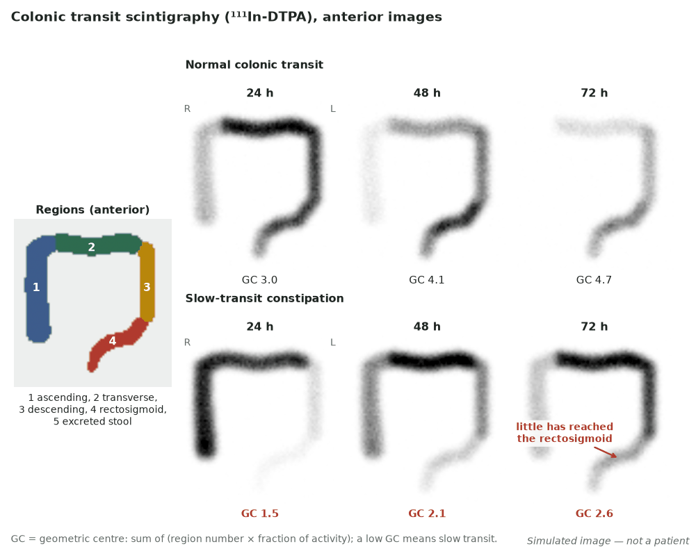
- Colon transit is imaged at 24, 48 and 72 h and summarised by the geometric centre (weighted mean across colonic segments; low = caecum, high = rectosigmoid/excreted).
- Reference geometric centres (seven-region method) are 2.0–7.0 at 24 h, 4.6–7.0 at 48 h and 6.0–7.0 at 72 h.
- Abnormal patterns: generalised slow transit (GC <4.1 at 48 h), colonic inertia (no progress beyond splenic flexure), and functional outlet obstruction (reaches rectosigmoid but not expelled).
- Small-bowel transit is interpretable only if liquid gastric emptying and colon transit are normal, else the report must be qualified.
Sources: SNMMI/EANM small-bowel and colon transit guideline 1.0, J Nucl Med 2013 (PMID 24092937)