Role of Nuclear Medicine in Movement Disorders
At a glance
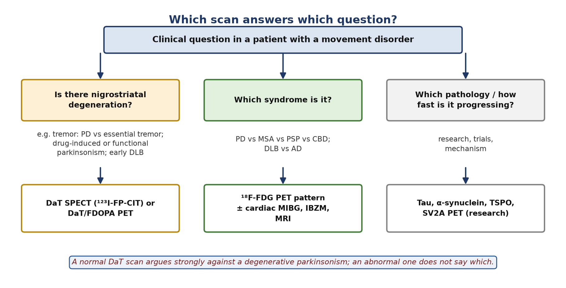
- Two questions. Presynaptic imaging (DaT SPECT/PET, FDOPA) answers is there nigrostriatal degeneration?; FDG PET patterns answer which syndrome?
- Normal DaT scan. Argues strongly against PD and other degenerative parkinsonisms (e.g. essential tremor, drug-induced or functional parkinsonism).
- Abnormal DaT scan. Confirms nigrostriatal loss but does not separate PD from MSA, PSP, CBD or DLB.
- FDG signatures. PD: relative lentiform/thalamic/cerebellar hypermetabolism; MSA: putamen, pons, cerebellum; PSP: midbrain and medial frontal; CBD: asymmetric frontoparietal; DLB: occipital with cingulate island.
- Cardiac MIBG. Reduced in PD and DLB (postganglionic denervation); usually preserved in MSA and PSP.
- Research tracers. Tau, α-synuclein, TSPO and SV2A PET are changing diagnosis but are not yet routine.
1. The basal ganglia circuit
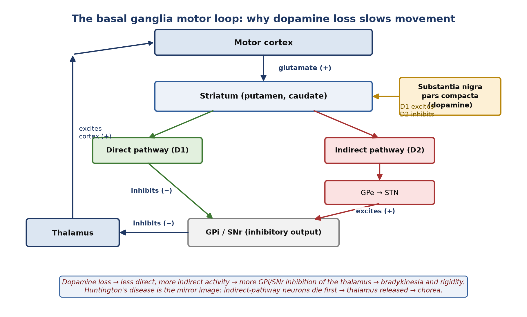
- The basal ganglia refine movement through a cortex → striatum → GPi/SNr → thalamus → cortex loop. Their output (GPi/SNr) is inhibitory by default.
- Direct pathway (D1 receptors): facilitates movement.
- Indirect pathway (D2 receptors, via GPe and STN): suppresses movement.
- Dopamine from the substantia nigra pars compacta excites the direct pathway and inhibits the indirect pathway, tipping the balance towards movement.
- Glutamate, GABA, acetylcholine, noradrenaline and serotonin act on the same circuit, which is why non-dopaminergic disease and drugs also cause movement disorders.
2. The clinical problem
- MDS criteria (2015): parkinsonism = bradykinesia plus rest tremor and/or rigidity. 'Clinically established' PD needs no absolute exclusions, at least two supportive criteria and no red flags; 'clinically probable' allows red flags only if counterbalanced by supportive criteria.
- Even so, clinical diagnosis is wrong in a meaningful minority, especially early; clinicopathological series put accuracy at roughly 80%.
| Feature | Idiopathic PD | Red flag for atypical parkinsonism |
|---|---|---|
| Onset | Usually asymmetric | Early symmetry: PSP, MSA |
| Levodopa response | Clear and sustained | Poor or short-lived |
| Autonomic failure | Later | Early and severe: MSA |
| Falls, gaze palsy | Late, if at all | Early falls, vertical gaze palsy: PSP |
| Cortical signs | Absent early | Apraxia, alien limb, cortical sensory loss: CBD |
| Hallucinations, cognition | Usually later | Early dementia and visual hallucinations: DLB |
| Parkinsonian tremor | Essential tremor | |
|---|---|---|
| Type | Rest tremor, 4–6 Hz, 'pill-rolling'; lessens with action | Postural/action tremor, 4–12 Hz; worse with movement |
| Distribution | Asymmetric, one-sided onset | Usually bilateral and symmetric |
| Company it keeps | Bradykinesia, rigidity | No bradykinesia; often familial; may improve with alcohol |
| DaT SPECT | Abnormal | Normal |
Diseases by the protein they accumulate
| Group | Protein | Disorders |
|---|---|---|
| Synucleinopathies | α-synuclein | PD, DLB, MSA |
| Tauopathies | 4-repeat tau | PSP, CBD |
| Polyglutamine disorders | Mutant huntingtin (and other proteins) | Huntington's disease, several spinocerebellar ataxias |
3. The tracers
| Target | SPECT | PET |
|---|---|---|
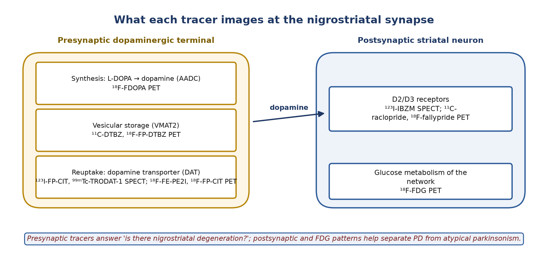
| Dopamine transporter (presynaptic) | ¹²³I-FP-CIT (ioflupane, DaTscan); ⁹⁹ᵐTc-TRODAT-1 | ¹⁸F-FP-CIT, ¹⁸F-FE-PE2I, ¹¹C-CFT |
| Dopamine synthesis (AADC) | — | ¹⁸F-FDOPA |
| Vesicular storage (VMAT2) | — | ¹¹C-DTBZ, ¹⁸F-FP-DTBZ |
| D2/D3 receptors (postsynaptic) | ¹²³I-IBZM | ¹¹C-raclopride, ¹⁸F-fallypride |
| Glucose metabolism | — | ¹⁸F-FDG |
| Cardiac sympathetic nerves | ¹²³I-MIBG | — |
| Research: tau / α-synuclein / microglia / synapses | — | ¹⁸F-PI-2620, ¹⁸F-florzolotau / ¹⁸F-ACI-12589 / TSPO ligands (¹¹C-PK11195) / SV2A (¹¹C-UCB-J) |
4. Protocols
| DaT SPECT (¹²³I-FP-CIT) | ¹⁸F-FDOPA PET | ¹⁸F-FDG brain PET | |
|---|---|---|---|
| Fasting | Not required | No amino-acid-containing food for 4 h | Fast at least 4–6 h; water allowed |
| Drugs | If clinically safe, stop drugs that bind DAT (e.g. cocaine, amphetamines, methylphenidate, bupropion, modafinil); antiparkinsonian drugs do not interfere significantly | Antiparkinsonian drugs do not significantly affect visual reading; consider stopping levodopa for ≥12 h | Avoid sedatives where possible; record all drugs |
| Premedication | Thyroid blockade ≥1 h before injection (e.g. iodide equivalent to 100 mg, or potassium perchlorate 400 mg) | Carbidopa 150 mg (or 2 mg/kg, max 150 mg) or entacapone 200 mg, 60–90 min before | None |
| Activity (adult) | 110–250 MBq (typically 185 MBq) | 185 MBq | 125–250 MBq (typically 150 MBq) for 3D PET; less on high-sensitivity digital systems |
| Imaging | SPECT 3–6 h after injection; keep a fixed time (e.g. 3 h) | Static or dynamic acquisition; striatal uptake over the first ~90 min is assessed | Quiet, dimly lit room during uptake; static scan at a fixed time 30–60 min after injection |
Table 1. Summary protocols (EANM/SNMMI guidelines). Follow local protocol and product information.
5. Reading a DaT scan
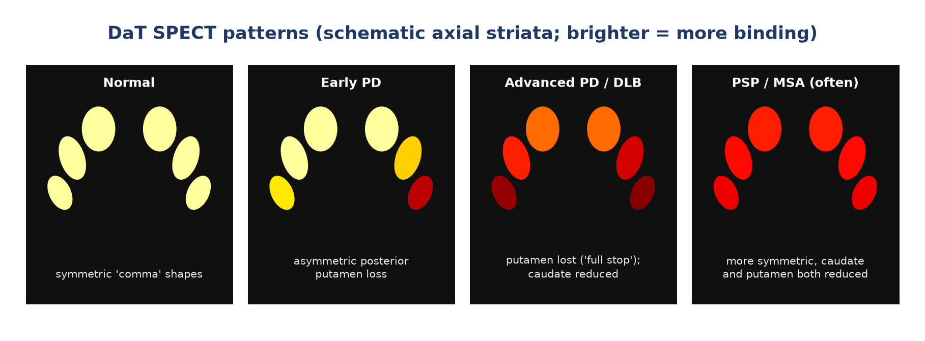
- Normal: symmetric comma- or crescent-shaped striatal uptake.
- Abnormal: loss begins in the posterior putamen, usually opposite the more affected side; the striatum becomes oval ('full stop' rather than 'comma').
- Semiquantification: specific binding ratios, putamen/caudate ratio and asymmetry index compared with an age-matched normal database add objectivity, especially in early or borderline cases.
- Visual reading remains the reference; quantification supports it.
Limitations
- An abnormal scan does not distinguish PD from MSA, PSP, CBD or DLB.
- A small minority of patients diagnosed clinically with PD have a normal scan (SWEDD: scans without evidence of dopaminergic deficit); most later prove not to have PD.
- DaT imaging is a diagnostic test, not a stand-alone measure of severity or progression.
Other dopaminergic and autonomic tests
- FDOPA PET: reads like DaT imaging (posterior putamen first) and is also used in trials.
- ¹²³I-IBZM SPECT (D2/D3): postsynaptic receptors are preserved or up-regulated in PD but reduced in MSA and PSP. Less standardised, used selectively.
- Cardiac ¹²³I-MIBG: the heart-to-mediastinum ratio is reduced in PD and DLB (postganglionic sympathetic denervation) and usually preserved in MSA and PSP. Cut-offs depend on the camera and collimator, so use the local normal range.
6. FDG PET patterns
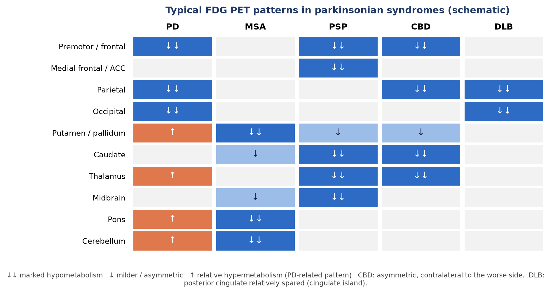
| Disease | Signature pattern |
|---|---|
| PD | PD-related pattern: premotor and parieto-occipital hypometabolism with relative hypermetabolism of pallidum/putamen, thalamus, pons and cerebellum |
| MSA | Hypometabolism of putamen (MSA-P) and/or pons and cerebellum (MSA-C) |
| PSP | Midbrain, medial frontal and anterior cingulate, caudate and thalamic hypometabolism |
| CBD / CBS | Asymmetric frontoparietal (especially inferior parietal), caudate and thalamic hypometabolism, contralateral to the worse side |
| DLB | Occipital and parietal hypometabolism with relative sparing of the posterior cingulate (cingulate island sign) |
7. Disease by disease
Parkinson's disease
- DaT, FDOPA and VMAT2 imaging show asymmetric, posterior-putamen-predominant presynaptic loss; D2 receptors are preserved.
- FDG shows the PD-related pattern; cardiac MIBG is often reduced.
- Non-dopaminergic PET (cholinergic, serotonergic, SV2A synaptic density) explains features such as falls, hallucinations and cognitive decline; research use.
Multiple system atrophy
- Early severe autonomic failure; MSA-P (parkinsonism) or MSA-C (cerebellar ataxia); poor levodopa response; MRI may show putaminal rim or 'hot-cross-bun' signs.
- DAT loss is more diffuse than in PD; FDG shows putaminal and/or pontocerebellar hypometabolism; cardiac MIBG usually preserved.
- α-synuclein PET (¹⁸F-ACI-12589) shows increased binding in the cerebellar white matter and middle cerebellar peduncles in MSA (research).
Progressive supranuclear palsy
- Early falls, vertical gaze palsy (slowed vertical saccades), axial rigidity, poor levodopa response.
- Marked, more symmetric striatal DAT loss including the caudate; FDG: midbrain and medial frontal/anterior cingulate hypometabolism.
- Second-generation tau PET (¹⁸F-PI-2620, ¹⁸F-florzolotau) shows 4R-tau in the basal ganglia, subthalamic nucleus, midbrain and dentate nucleus.
Corticobasal degeneration / syndrome
- Asymmetric rigidity or dystonia, apraxia, cortical sensory loss, alien-limb phenomenon. The syndrome can be caused by CBD, PSP, Alzheimer or FTLD pathology.
- FDG: asymmetric frontoparietal hypometabolism opposite the clinically worse side; DAT loss is asymmetric and does not separate CBD from PD on its own.
Dementia with Lewy bodies and PD dementia
- Reduced striatal DAT uptake and reduced cardiac MIBG uptake are indicative biomarkers of DLB (4th consensus criteria).
- FDG: occipital hypometabolism with a cingulate island sign favours DLB over Alzheimer disease; amyloid PET is often positive as co-pathology and is not specific.
Ataxias
- Hereditary (spinocerebellar ataxias, Friedreich ataxia), acquired (alcohol, autoimmune/paraneoplastic, vitamin E deficiency, hypothyroidism) and sporadic (including MSA-C). Treatable acquired causes must be excluded first.
- FDG: cerebellar hypometabolism in most spinocerebellar ataxias and MSA-C; added pontine/brainstem involvement in MSA-C, SCA2 and SCA3; cortical or striatal involvement in some (e.g. SCA17, DRPLA).
Huntington's disease
- Autosomal dominant CAG-repeat expansion in HTT; chorea, cognitive decline and psychiatric features.
- Indirect-pathway (D2) striatal neurons are lost first, releasing the thalamus (Figure 1).
- FDG: caudate and putaminal hypometabolism, which begins before symptoms in gene carriers; ¹¹C-raclopride shows reduced striatal D2 binding, also before symptoms.
- PDE10A PET and TSPO (microglial) PET are research markers of progression.
Summary
- Use DaT SPECT/PET or FDOPA to answer whether there is nigrostriatal degeneration.
- A normal scan argues strongly against degenerative parkinsonism; an abnormal scan does not name the disease.
- Use FDG PET patterns, cardiac MIBG and IBZM to separate PD from MSA, PSP, CBD and DLB.
- In DLB, reduced DAT and reduced MIBG are indicative biomarkers.
- Tau, α-synuclein, TSPO and SV2A PET are research tools that are moving towards the clinic.
Test yourself
5 quick questions. Pick an answer to see the explanation.
1. A 68-year-old has a bilateral postural tremor for 10 years, no bradykinesia, and improvement with alcohol. DaT SPECT is most likely to show:
2. An abnormal DaT scan in a patient with parkinsonism:
3. Which FDG PET pattern best fits PSP?
4. Cardiac MIBG uptake is typically reduced in:
5. Before ¹⁸F-FDOPA PET, carbidopa is given to:
References
- Postuma RB, Berg D, Stern M, et al. MDS clinical diagnostic criteria for Parkinson's disease. Mov Disord. 2015;30(12):1591-601.
- Morbelli S, Esposito G, Arbizu J, et al. EANM practice guideline/SNMMI procedure standard for dopaminergic imaging in Parkinsonian syndromes 1.0. Eur J Nucl Med Mol Imaging. 2020;47(8):1885-912.
- Guedj E, Varrone A, Boellaard R, et al. EANM procedure guidelines for brain PET imaging using [18F]FDG, version 3. Eur J Nucl Med Mol Imaging. 2022;49(2):632-51.
- Rizzo G, Copetti M, Arcuti S, et al. Accuracy of clinical diagnosis of Parkinson disease: a systematic review and meta-analysis. Neurology. 2016;86(6):566-76.
- McKeith IG, Boeve BF, Dickson DW, et al. Diagnosis and management of dementia with Lewy bodies: fourth consensus report of the DLB Consortium. Neurology. 2017;89(1):88-100.
- Höglinger GU, Respondek G, Stamelou M, et al. Clinical diagnosis of progressive supranuclear palsy: the Movement Disorder Society criteria. Mov Disord. 2017;32(6):853-64.
Practise with a case
- Slowness and falls in a 66-year-old taking risperidone · Intermediate