Dopamine Transporter Imaging (DaTSCAN)
¹²³I-FP-CIT (ioflupane, DaTSCAN) SPECT images the presynaptic dopamine transporter in the striatum. It reliably separates degenerative parkinsonism (Parkinson’s disease, atypical syndromes) from conditions with intact dopaminergic terminals — essential tremor, drug-induced and psychogenic parkinsonism — and is an indicative biomarker for dementia with Lewy bodies (sensitivity ≈78%, specificity ≈90%). It cannot, however, distinguish Parkinson’s disease from atypical parkinsonian syndromes.
In Parkinson’s disease and related disorders the nigrostriatal dopaminergic neurons degenerate, reducing striatal DAT density. DaTSCAN visualises this loss, typically beginning in the putamen and often asymmetric.
When to image
- Clinically uncertain tremor or parkinsonism — degenerative vs essential/drug-induced.
- Supporting a diagnosis of dementia with Lewy bodies.
- Evaluating suspected psychogenic parkinsonism.
- Not required when the clinical diagnosis of Parkinson’s disease is clear.
How to read it
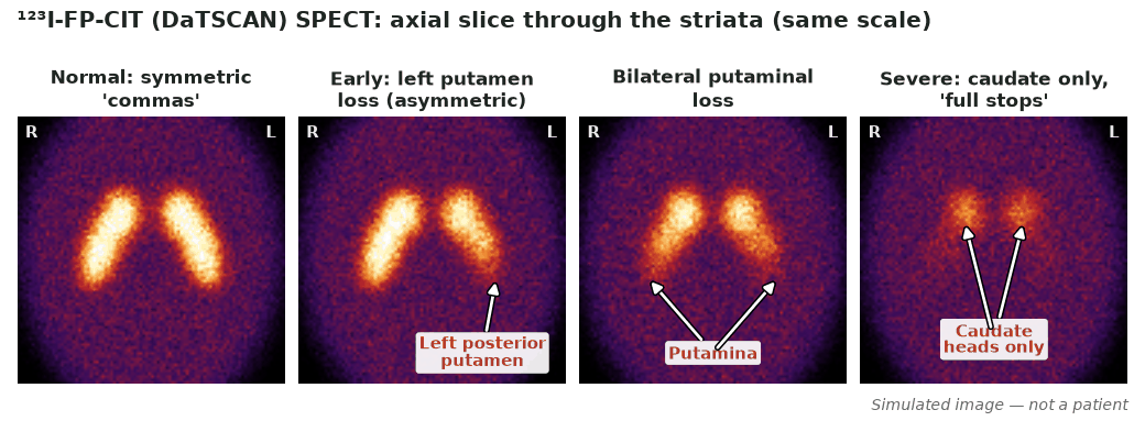
- Normal: symmetric “comma/crescent” striatal uptake.
- Abnormal: reduced uptake, usually starting in the putamen and often asymmetric (“full-stop” appearance).
- A clearly normal scan argues against a presynaptic dopaminergic disorder.
- Uptake loss does not distinguish PD from MSA/PSP/CBD.
Protocol
- Thyroid blockade (potassium iodide or perchlorate) at least 1 h before injection to reduce thyroid exposure to free ¹²³I.
- Image ~3–6 h after injection with high-resolution SPECT (± CT).
- Semi-quantitative striatal binding ratios support visual assessment.
Diagnostic performance
For distinguishing degenerative parkinsonism from essential tremor, sensitivity and specificity are high (≈95% and ≈93% on blinded consensus reading in the multicentre Benamer study). For dementia with Lewy bodies, a phase III study reported sensitivity ≈78% and specificity ≈90% versus non-DLB dementia.
Pitfalls
- Some drugs (e.g. certain stimulants, cocaine) affect DAT binding — review medications.
- Cannot differentiate Parkinson’s disease from atypical parkinsonian syndromes.
- Age-related DAT decline (≈5.5–6% per decade) must be accounted for — compare with age-matched normal databases.
Evidence & guidelines
- McKeith et al. (Lancet Neurology, 2007): DaTSCAN sensitivity ≈78%, specificity ≈90% for DLB.
- Reduced striatal DAT uptake is an indicative biomarker in DLB consensus criteria.
- The EANM/SNMMI dopaminergic imaging guideline (Morbelli et al., 2020) covers acquisition and interpretation.
In depth
- Among SPECT dopamine transporter tracers, only [¹²³I]FP-CIT (ioflupane) is approved by both the FDA and EMA; the typical adult activity is 110–250 MBq (usually 185 MBq), and [¹⁸F]fluorodopa PET is a clinical alternative.
- Thyroid blockade against free ¹²³I is given ≥1 h before injection (potassium iodide/Lugol’s ≈100 mg iodide, potassium perchlorate 400 mg, or sodium perchlorate 600 mg).
- Image 3–6 h after [¹²³I]FP-CIT (18–24 h for [¹²³I]β-CIT); each centre should keep a fixed start time for comparability.
- Stop interfering drugs for ≥5 half-lives — cocaine, amphetamines, methylphenidate and modafinil are high-affinity DAT blockers that markedly reduce binding.
- SSRIs may increase striatal binding (~20% for β-CIT, ~10% for FP-CIT); standard anti-parkinsonian drugs, neuroleptics and cholinesterase inhibitors do not significantly interfere.
- Normal striata are comma-shaped and symmetric; abnormal scans show reduced oval/circular striata, with early Parkinson’s disease affecting the posterior putamen contralateral to symptoms first (‘dot’ shape).
- The specific binding ratio (SBR, striatal target-to-background) is the commonest semiquantitative measure but is not a direct measure of DAT density; normal ageing reduces SBR ~5.5–6% per decade.
- DAT SPECT helps separate Alzheimer’s disease (typically normal) from dementia with Lewy bodies (reduced), but about 10% of pathologically confirmed DLB cases have a normal scan at clinical diagnosis, which may become abnormal after 1.5–2 years.
- Binding is normal in essential tremor, drug-induced and psychogenic parkinsonism, and DAT SPECT cannot distinguish Parkinson’s disease/DLB from PSP, CBD or the putaminal MSA variant.
- Acquisition: LEHR/LEUHR collimators, ≥128×128 matrix, 360° with ~3° steps, >1.5 million counts (FP-CIT), reconstructed resolution ≤10 mm FWHM.
Sources: EANM/SNMMI dopaminergic imaging guideline 2020 (PMID 32388612)
- Slowness and falls in a 66-year-old taking risperidone · Intermediate