Brain Perfusion SPECT
Brain perfusion SPECT with ⁹⁹ᵐTc-HMPAO or ⁹⁹ᵐTc-ECD maps regional cerebral blood flow. Its enduring roles are localising the epileptogenic focus (ictal injection), assessing cerebrovascular reserve (with acetazolamide), supporting a dementia diagnosis where FDG PET is unavailable, and confirming brain death. PET has largely superseded it for dementia and tumours, but ictal SPECT and brain-death confirmation remain distinctive strengths.
HMPAO and ECD are lipophilic tracers that cross the blood–brain barrier and are trapped intracellularly in proportion to perfusion at the instant of injection — a “frozen” snapshot. This fixation is what allows a tracer injected during a seizure to be imaged afterwards, capturing ictal blood flow.
Clinical uses
- Ictal seizure localisation (tracer injected at seizure onset).
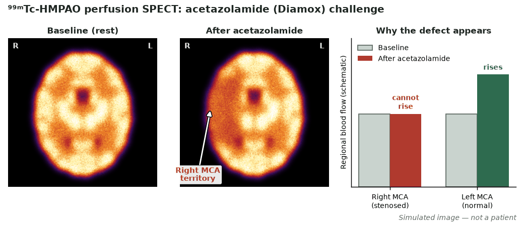
- Cerebrovascular reserve before revascularisation, using an acetazolamide (Diamox) challenge.
- Dementia support where FDG PET is unavailable (temporoparietal hypoperfusion in Alzheimer’s).
- Ancillary confirmation of brain death (perfusion agents).
Protocol
- Inject in a quiet, dimly lit room to standardise cortical activity (interictal studies).
- For ictal SPECT, inject at seizure onset under EEG/video monitoring, then image once the patient is stable.
- Acetazolamide challenge compares baseline and post-vasodilator perfusion to reveal exhausted reserve.
- SPECT (± CT) acquisition with iterative reconstruction.
How to read it
- Alzheimer’s: temporoparietal and posterior cingulate hypoperfusion.
- Frontotemporal dementia: frontal and anterior temporal hypoperfusion.
- DLB: occipital hypoperfusion in addition to posterior changes.
- Epilepsy: ictal hyperperfusion and interictal hypoperfusion at the focus.
Strengths & limitations
- Ictal snapshot is a unique strength for epilepsy.
- Widely available and lower cost than PET.
- Lower spatial resolution and quantitation than FDG PET.
- Interictal perfusion SPECT alone is insensitive for seizure localisation.
Pitfalls
- Delayed ictal injection captures propagation rather than onset.
- Atrophy and partial-volume effects reduce apparent perfusion.
- In subacute stroke ECD, unlike HMPAO, does not show luxury perfusion (reperfusion hyperaemia) — the two tracers are not interchangeable.
Evidence & guidelines
- The EANM procedure guideline for brain perfusion SPECT (Kapucu et al., 2009) covers technique and interpretation.
- Ictal SPECT (often with SISCOM co-registration) is an established tool in epilepsy surgery work-up.
- PET has superseded perfusion SPECT for most dementia and tumour indications.
In depth
- ⁹⁹ᵐTc-HMPAO and ⁹⁹ᵐTc-ECD are lipophilic, brain-specific perfusion tracers that cross the blood–brain barrier and are retained; unlike ⁹⁹ᵐTc-DTPA, their interpretation depends much less on bolus quality and they allow delayed imaging.
- Because HMPAO and ECD are retained in the brain, delayed planar images (about 20 min after injection) with or without SPECT can still give a definitive result when the angiographic bolus is poor.
- Static planar images are acquired in anterior, both lateral and, if possible, posterior projections (about 500,000–1,000,000 counts per view with non-specific agents). Brain-specific agents show cerebral and cerebellar perfusion, and SPECT better demonstrates the posterior fossa and brainstem.
- The posterior fossa can be assessed with brain-specific agents on lateral and posterior views and, better, with SPECT; anterior-only DTPA flow imaging cannot evaluate it.
- An acetazolamide (Diamox) challenge compares baseline and post-vasodilator perfusion SPECT to reveal exhausted cerebrovascular reserve.
- Brain-specific agents (HMPAO, ECD) are preferred by many centres because interpretation depends less on the bolus and delayed images are usually definitive, although the SNM guideline notes no clear evidence that they are more accurate than non-specific agents such as DTPA.
Sources: SNM practice guideline for brain death scintigraphy 2.0 (PMID 22743146) · EANM brain perfusion SPECT guideline v2 2009 (PMID 19838703)
Spotted an error or something out of date? Report an error