Patient Preparation
Correct preparation is the commonest avoidable determinant of a diagnostic study. It is study-specific: fasting and glucose control for FDG oncology, opposite dietary regimens for cardiac FDG, caffeine abstinence before vasodilator stress, a low-iodine diet and TSH stimulation before radioiodine, thyroid blockade and a drug review before mIBG and DaT imaging, and hydration with voiding for renally excreted tracers.
Preparation raises target uptake, suppresses competing physiological uptake and lowers the radiation dose. Most errors cannot be corrected once the tracer is in the patient, so the checks — identity, indication, pregnancy and breastfeeding status, recent iodine loads, interfering drugs and blood glucose — are made before the dose is drawn up.
- FDG oncology: fast ≥4 h (EANM 2015; SNMMI 4–6 h), water encouraged (about 1 L in the 2 h before injection); inject if plasma glucose is <11 mmol/L (about 200 mg/dL); uptake 60 min (55–75 min acceptable).
- Insulin and FDG: inject ≥4 h after subcutaneous rapid-acting insulin and ≥6 h after short-acting insulin; no strenuous exercise for ≥6 h, preferably 24 h.
- Cardiac sarcoidosis FDG: ≥2 high-fat (>35 g), low-carbohydrate (<3 g) meals the day before, then fast ≥4–12 h; or fast >18 h; optional heparin 50 IU/kg IV about 15 min before FDG.
- Vasodilator stress MPI: no caffeine or other methylxanthines for ≥12 h; stop dipyridamole ≥24 h before adenosine or regadenoson.
- Radioiodine for thyroid cancer: low-iodine diet (≤50 µg/day) for about 1–2 weeks; TSH >30 mIU/L after LT4 withdrawal for 3–4 weeks, or rhTSH 0.9 mg IM twice 24 h apart with ¹³¹I 24 h after the second injection.
- After IV iodinated contrast: ESUR v10 advises 2 months before radioiodine therapy or thyroid imaging; ATA 2015 accepts 4–8 weeks; ACR suggests 3–4 weeks (hyperthyroid) or 6 weeks (hypothyroid).
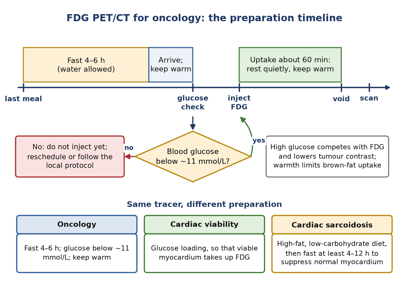
FDG PET/CT for oncology
- Fast for at least 4 h (EANM v2.0; the SNMMI guideline says 4–6 h). Plain water is encouraged — about 1 L in the 2 h before injection — to dilute urinary FDG, reduce artefacts and lower the bladder dose.
- Check plasma glucose before injection. Below 11 mmol/L (about 200 mg/dL) the study can go ahead; at or above it, reschedule or follow the local diabetic protocol. Research protocols often set a stricter ceiling of 7–8.3 mmol/L.
- Do not give insulin just before FDG to bring glucose down: insulin drives FDG into muscle and fat. Rapid-acting insulin peaks at about 60 min and acts for 2–4 h, so FDG is given at least 4 h after it (6 h after short-acting insulin). Patients on tablets are best scanned in the late morning.
- Avoid strenuous exercise for at least 6 h, preferably 24 h, to prevent muscle uptake. Keep the patient warm from 30–60 min before injection until the scan to limit brown-fat uptake.
- Keep the uptake time at 60 min (acceptable range 55–75 min). For response assessment, repeat the previous interval to within 10 min so that SUVs remain comparable. Void immediately before imaging.
Cardiac and brain studies
- Cardiac sarcoidosis FDG aims to switch normal myocardium from glucose to fatty-acid use: at least two high-fat (>35 g), low-carbohydrate (<3 g) meals the day before and a fast of at least 4–12 h, or a fast of more than 18 h. Heparin 50 IU/kg IV about 15 min before FDG is an optional adjunct of uncertain value (SNMMI/ASNC 2017). See cardiac sarcoidosis.
- FDG viability imaging needs the opposite: glucose loading, with insulin as required, so that viable myocardium takes up FDG. See viability.
- Vasodilator stress perfusion imaging: no caffeine or other methylxanthines for at least 12 h (long-acting methylxanthines for five half-lives), because they block adenosine receptors and blunt hyperaemia. Stop dipyridamole at least 24 h before adenosine or regadenoson. Anti-anginal drugs are withheld for 3–5 half-lives when the question is diagnostic, by agreement with the referrer (EANM 2015).
- DaT SPECT: stop drugs that block the dopamine transporter for about five half-lives — cocaine 2 days, amphetamines and methylphenidate 7 days, modafinil 3 days, bupropion 8 days. Levodopa, dopamine agonists and MAO-B inhibitors need not be stopped. Give thyroid blockade (for example 100 mg iodide or 400 mg potassium perchlorate) at least 1 h before injection (EANM/SNMMI 2020). See DaT imaging.
Radioiodine and mIBG
- Differentiated thyroid cancer: a low-iodine diet (≤50 µg/day) for about 1–2 weeks, and TSH above 30 mIU/L, either by withdrawing levothyroxine for 3–4 weeks (liothyronine can bridge the early weeks but is stopped for at least 2 weeks) or with rhTSH 0.9 mg IM on two occasions 24 h apart (ATA 2015). Radioiodine is given 24 h after the second rhTSH injection; a diagnostic scan follows 48 h later and serum thyroglobulin is taken 72 h after the last injection (Thyrogen label).
- Ask about iodine loads: iodinated contrast, amiodarone and iodine-containing supplements. ESUR v10 advises no iodinated contrast for at least 2 months before radioiodine therapy; the ATA accepts 4–8 weeks, by which time most of the iodine has cleared. Urinary iodine can be measured when contamination is suspected. See contrast in hybrid imaging.
- Before radioiodine therapy, confirm a negative pregnancy test and that breastfeeding (or pumping) stopped at least 3 months earlier (ATA 2015); lactating breast tissue concentrates iodide. See pregnancy & breastfeeding.
- mIBG: block the thyroid from the day before injection and continue for 1–2 days after ¹²³I-mIBG or 2–3 days after ¹³¹I-mIBG, for example potassium iodide 130 mg daily; potassium perchlorate 400 mg is the alternative for iodine allergy or same-day starts (EANM 2010).
- mIBG interfering drugs and withdrawal times (EANM 2010): labetalol 72 h; reserpine 48 h; most calcium-channel blockers 48 h; sympathomimetics 24 h (nasal decongestants 48 h); tricyclic antidepressants 24–48 h. Amiodarone cannot practically be stopped. Stopping α- or β-blockade is agreed with the referrer. See mIBG imaging.
Other common studies
- Gastric emptying: overnight fast (at least 6 h); stop prokinetics (metoclopramide, domperidone, erythromycin) and opiates 2 days before; fasting glucose should be below 275 mg/dL (about 15 mmol/L), because hyperglycaemia itself slows emptying; no smoking on the morning of the test (Abell 2008). See gastric emptying.
- PSMA PET: no fasting and no need to stop medication. Drink water; for renally excreted ligands (⁶⁸Ga-PSMA-11, ¹⁸F-DCFPyL) furosemide 20 mg IV or 1 L of oral fluid during uptake helps to clear urinary activity; this is not needed for ¹⁸F-PSMA-1007. Void just before imaging (EANM/SNMMI 2023).
- Renal and bone studies: good hydration and frequent voiding speed washout and reduce bladder and gonadal dose.
Checks before every administration
- Confirm identity, the request and its justification, and the radiopharmaceutical and activity against the request.
- Ask about pregnancy and breastfeeding in everyone of childbearing potential; UK employers must have a written procedure for this under IR(ME)R, with a stated age range (ARSAC gives 12–55 years as an example).
- Check renal function only where it changes management: before iodinated contrast and before renally cleared therapies.
Pitfalls
- Hyperglycaemia or recent insulin lowers tumour FDG uptake and raises muscle uptake.
- Iodinated contrast, amiodarone or iodine supplements block radioiodine uptake for weeks.
- An unrecognised interfering drug — labetalol before mIBG, amphetamines or methylphenidate before DaT SPECT — causes false-negative uptake.
- Inadequate dietary suppression leaves diffuse myocardial FDG uptake that cannot be read for sarcoidosis.
- Caffeine within 12 h of adenosine or regadenoson blunts hyperaemia and can hide ischaemia.
In depth
- EANM FDG v2.0 scales activity to body weight and bed time: FDG (MBq) = 14 × weight (kg) ÷ minutes per bed position when bed overlap is ≤30%, and 7 × weight ÷ minutes per bed when overlap is >30%; a quadratic scheme referenced to 75 kg is offered for heavy patients.
- Positive oral contrast should be diluted if SUVs are to be quantified; concentrated positive agents cause attenuation-correction errors in the bowel.
- SSRIs increase striatal [¹²³I]FP-CIT binding by about 10%. This matters for research quantification, not for clinical visual reads, so they are not stopped.
- The ATA recommendation for TSH >30 mIU/L is graded weak: the optimum level for remnant ablation is uncertain. Liothyronine bridging is used only when levothyroxine is withdrawn for 4 weeks or more.
- For gastric emptying, insulin-treated diabetic patients take about half their usual dose with the test meal, and premenopausal women are preferably studied in days 1–10 of the cycle because hormonal phase alters emptying.
- Dipyridamole side effects are reversed with aminophylline or theophylline 125–250 mg IV — the same methylxanthine effect that makes caffeine abstinence necessary.
- PSMA uptake time is about 60 min (acceptable 50–100 min) for most ligands but 90–120 min for ¹⁸F-PSMA-1007, whose low urinary excretion is also why it needs no furosemide.
Sources: EANM FDG PET/CT v2.0 (Boellaard 2015, PMID 25452219) · EANM/SNMMI dopaminergic imaging 2020 (PMID 32388612) · ATA 2015 (PMID 26462967) · Abell 2008 gastric emptying consensus (PMID 18287197) · EANM MPI 2015 (PMID 26290421) · EANM/SNMMI PSMA 2.0 (PMID 36604326)
Sources
- Boellaard R, Delgado-Bolton R, Oyen WJG, et al. FDG PET/CT: EANM procedure guidelines for tumour imaging: version 2.0. Eur J Nucl Med Mol Imaging. 2015;42:328–354.
- Chareonthaitawee P, Beanlands RS, Chen W, et al. Joint SNMMI–ASNC expert consensus document on the role of ¹⁸F-FDG PET/CT in cardiac sarcoid detection and therapy monitoring. J Nucl Cardiol. 2017;24:1741–1758.
- Verberne HJ, Acampa W, Anagnostopoulos C, et al. EANM procedural guidelines for radionuclide myocardial perfusion imaging with SPECT and SPECT/CT: 2015 revision. Eur J Nucl Med Mol Imaging. 2015;42:1929–1940.
- Haugen BR, Alexander EK, Bible KC, et al. 2015 American Thyroid Association management guidelines for adult patients with thyroid nodules and differentiated thyroid cancer. Thyroid. 2016;26:1–133.
- Bombardieri E, Giammarile F, Aktolun C, et al. ¹³¹I/¹²³I-metaiodobenzylguanidine (mIBG) scintigraphy: procedure guidelines for tumour imaging. Eur J Nucl Med Mol Imaging. 2010;37:2436–2446.
- Morbelli S, Esposito G, Arbizu J, et al. EANM practice guideline/SNMMI procedure standard for dopaminergic imaging in Parkinsonian syndromes 1.0. Eur J Nucl Med Mol Imaging. 2020;47:1885–1912.
- Abell TL, Camilleri M, Donohoe K, et al. Consensus recommendations for gastric emptying scintigraphy: a joint report of the American Neurogastroenterology and Motility Society and the Society of Nuclear Medicine. J Nucl Med Technol. 2008;36:44–54.
- Fendler WP, Eiber M, Beheshti M, et al. PSMA PET/CT: joint EANM procedure guideline/SNMMI procedure standard for prostate cancer imaging 2.0. Eur J Nucl Med Mol Imaging. 2023;50:1466–1486.
- European Society of Urogenital Radiology Contrast Media Safety Committee. ESUR Guidelines on Contrast Agents, version 10.0. 2018.
- Thyrogen (thyrotropin alfa for injection): US prescribing information. Revised 2023.